


Please Leave Us A Message
Privacy statement: Your privacy is very important to Us. Our company promises not to disclose your personal information to any external company with out your explicit permission.

 November 17, 2023
Honey is an ancient natural wound-healing agent and has been reintroduced to modern clinical wound care as it has various bioactivities. In this study, honey was incorporated into an alginate/PVA-based electrospun nanofibrous membrane to develop an efficient wound dressing material. The morphology and chemical composition of the nanofibrous membrane were observed by scanning electron microscopy and characterized via Fourier transform infrared spectroscopy, respectively, demonstrating that honey was successfully introduced to the nanofibers. The nanofibrous membranes with increasing honey content showed enhanced antioxidant activity, suggesting the ability to control the overproduction of reactive oxygen species. Disc diffusion assay and dynamic contact assay proved the antibacterial activity of the honey loaded nanofibers towards Gram-positive bacterium (Staphylococcus aureus) and Gram-negative bacterium (Escherichia coli). The cytotoxicity assay illustrated the non-cytotoxicity and biocompatibility of the nanofibrous membranes. Therefore, the developed honey/alginate/PVA nanofibrous membranes are promising for wound dressings.
Skin plays an important role in protecting the body from external environmental interferences such as pathogens and chemicals (Chua et al., 2016). Once the structure or function of skin is defective, the body is susceptible to microbial invasion and wound infection, which delays wound healing and can even be life-threatening (Unnithan, Gnanasekaran, Sathishkumar, Lee, & Kim, 2014). Wound dressing is an efficient and common method to promote wound healing. Conventional dressings, such as cotton and gauze, have the advantages of low cost and high absorption capacity. However, they play only a passive role in the healing process by simply isolating the wound from contaminations (Mele, 2016; Pilehvar-Soltanahmadi et al., 2018). Moreover, the dehydration of, and enhanced adhesion on, the wound as a result of conventional dressings, cause discomfort and pain to the patient as well as delaying wound healing (Mayet et al., 2014). An ideal wound dressing, on one hand, should have a corresponding structure that prevents microbial invasion and allows gaseous exchange (Jayakumar, Prabaharan, Sudheesh Kumar, Nair, & Tamura, 2011). On the other hand, the dressing materials should be biocompatible, absorb excess exudates, and possess bioactive properties to promote wound healing, such as anti-bacterial behavior and antioxidant potential (Chhatri et al., 2011; Naseri-Nosar & Ziora, 2018).
Various constructions of wound dressings have been explored to facilitate wound healing, such as sponges, hydrogels, hydrocolloids, and films (Simões et al., 2018). Among these, electrospun nanofibrous membrane possesses a three-dimensional supportive structure, small pore size, and high surface-to-volume ratio, which has been reported to show great potential as a wound dressing (Abdelgawad, Hudson, & Rojas, 2014). The three-dimensional supportive structure could mimic the structure of the natural extracellular matrix, which is conducive to cell growth, adhesion and proliferation (Zhang, Oh et al., 2017). The small pore size and high porosity of nanofibrous mat could facilitate gaseous exchange and bacterial isolation during wound repair (Chui, Mouthuy, & Ye, 2018). The high surface-to-volume ratio of nanofibers has been proved to be beneficial for the loading and delivery of drugs for wound recovery (Sill & Recum, 2008; Zhang, Lim, Ramakrishna, & Huang, 2005).
Numerous materials have been used as electrospinning materials, among which natural polymers exhibited various advantages for wound repair application, such as hydrophilicity, non-toxicity and assistance for cell adhesion and proliferation (Hsu et al., 2004). Alginate, as a natural polymer, is an anionic polysaccharide which has excellent biocompatibility and biodegradability (Coşkun et al., 2014). Moreover, alginate could efficiently absorb excess exudate and provide a moist environment during the wound healing process due to its high hydrophilicity (Coşkun et al., 2014; Summa et al., 2018). However, pure alginate is difficult to electrospin because of the high electrical conductivity, high surface tension (Xiao & Lim, 2018) and lack of chain entanglements of its aqueous solution (Li et al., 2013). Therefore, synthetic polymers such as polyvinyl alcohol (PVA), were added to increase the electrospinnability as well as the mechanical strength of alginate (Shen & Hsieh, 2014), while PVA was also identified as a favorable wound dressing material (Fu et al., 2016; Zhou et al., 2008).
To obtain a better healing effect, nanofibrous dressings incorporated with antibacterial agents, such as silver nanoparticles, metal oxide, and antibiotics, have recently been studied (Liu et al., 2018; Mokhena & Luyt, 2017; Shalumon et al., 2011). Honey, because of its antibacterial (Martinotti & Ranzato, 2018), anti-inflammatory, and antioxidant (Bertoncelj, Doberšek, Jamnik, & Golob, 2007) properties, has been used in wound care dating back to 2000 BCE (Minden-Birkenmaier & Bowlin, 2018). Honey has also been reported to show little toxicity on fibroblasts and to increase the rate of re-epithelialization (Ranzato, Martinotti, & Burlando, 2012). Ranzato et al. demonstrated that honey efficiently facilitated wound closure (Ranzato, Martinotti, & Burlando, 2013). Recently, honey has been incorporated in different polymers via electrospinning, such as silk fibroin, PVA and chitosan (Sarhan & Azzazy, 2015; Sarkar, Ghosh, Barui, & Datta, 2018; Yang et al., 2017).

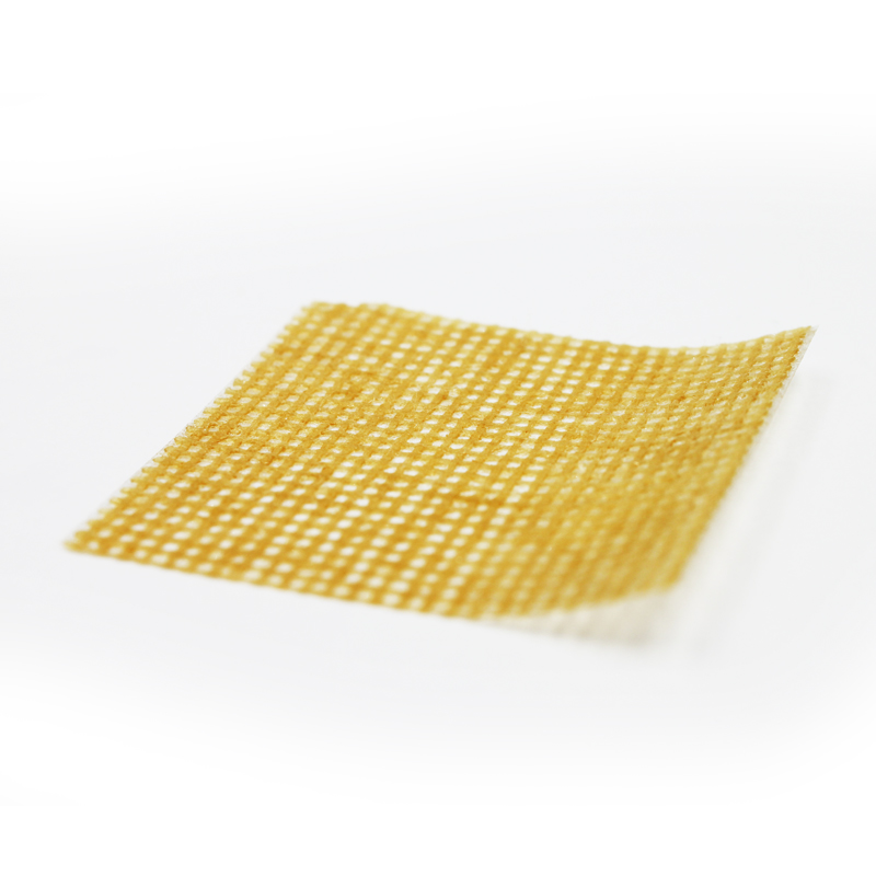
Figure 1. Honey/SA/PVA nanofibers fabricated by electrospinning. (a) Schematic illustration
of the solution preparation and electrospinning process. (b) Photograph of honey/SA/PVA
nanofiber membrane.

Figure 2. Water absorption (a) and weight loss (b) of the honey/SA/PVA nanofiber membranes
with varying honey content: 0%, 5%, 10%, 15%, and 20%. Results are mean ± SD (n=3).

Figure 3. Antioxidant activity of the honey/SA/PVA nanofiber membranes. (a) Photographs of
DPPH solutions after reaction with nanofiber membranes containing different honey content
(0%, 5%, 10%, 15%, and 20%) for 9 h. (b) DPPH radical scavenging activity of nanofibers
incorporated with different honey content (0%, 5%, 10%, 15%, and 20%). Results are mean ±
SD (n=3).

Figure 4. Antibacterial activity of the honey/SA/PVA nanofiber nanofibrous membranes with
varying honey content (0%, 5%, 10%, 15%, and 20%) evaluated by disc diffusion assay. (a-b)
Photographs of the inhibition zone against E. coli (a) and S. aureus (b). (c-d) Size of the
inhibition zone against E. coli (c) and S. aureus (d). Results are mean ± SD (n=3).
References
1.Tang Y, Lan X, Liang C, et al. Honey loaded alginate/PVA nanofibrous membrane as potential bioactive wound dressing[J]. Carbohydrate polymers, 2019, 219: 113-120.
Privacy statement: Your privacy is very important to Us. Our company promises not to disclose your personal information to any external company with out your explicit permission.
Fill in more information so that we can get in touch with you faster
Privacy statement: Your privacy is very important to Us. Our company promises not to disclose your personal information to any external company with out your explicit permission.